
Figure 3. Effects of SWNTs and METH on extracellular dopamine, dopamine oxidation and dopamine transporter (DAT) binding and function.
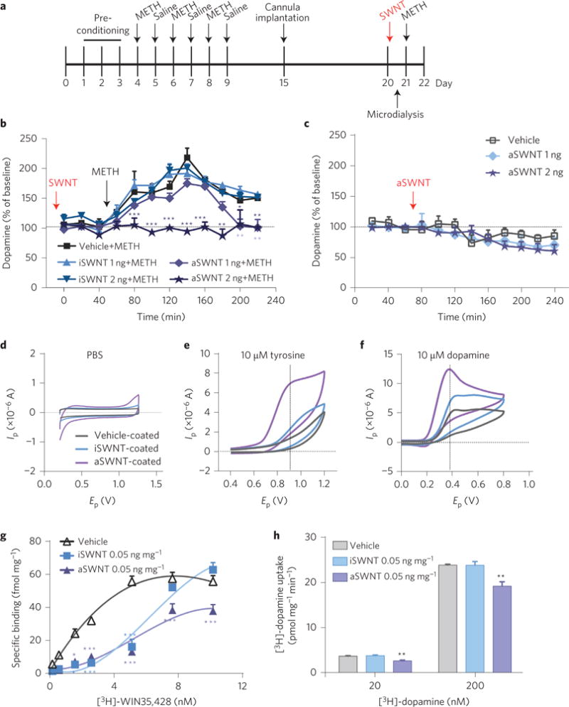
a, Timeline for the microdialysis experiment. b, Pretreatment with aSWNTs (1 or 2 ng by i.c.v. microinjection, n = 12 mice per group) dose-dependently attenuated METH-enhanced extracellular dopamine in the striatum. Two-way ANOVA with repeated measurements over time revealed a statistically significant SWNT treatment main effect (F4,55 = 43.5, p < 0.001) and time main effect (F11,605 = 76.6, p < 0.001). c, aSWNTs (1 or 2 ng by i.c.v. microinjection, n = 8 mice per group) alone did not significantly alter the basal levels of extracellular dopamine in the ventral striatum compared with the vehicle control group (n = 7 mice per group) (F2,20 = 0.45, p > 0.05). d–f, Representative fast-cyclic voltammograms, illustrating that SWNT-coated electrodes detected much larger tyrosine (10 μM) or dopamine (10 μM) oxidation currents than uncoated electrodes. Ip, oxidation current; Ep, applied voltage. g, Incubation of striatal tissue with aSWNTs (50 μg mg−1 tissue) inhibited [3H]-WIN35,428 binding to DAT in the striatal membrane preparations, and shifted the [3H]-WIN35,428 binding curves to the right (the maximal number of DAT binding sites Bmax = 78.23 ± 7.90 pmol mg−1; the equilibrium dissociation constant kd = 3.02 ± 0.81 nM). h, Incubation with aSWNTs (50 μg mg−1 tissue) also reduced re-uptake of [3H]-dopamine in striatal synaptosomes (20 nM: F2,9 = 19.09, p < 0.001; 200 nM: F2,9 = 13.83, p < 0.01, n = 4 mice per group). The maximal velocity of dopamine uptake Vmax = 6.50 ± 0.43 fmol mg−1 min−1; the dopamine concentration at 1/2 Vmax km = 336.3 ± 68.4 nM−1. The error bars indicate the s.e.m. of the means from 7–12 animals in each group (b,c) or from 4 different striatal samples (g,h). Each striatal sample was replicated three times. *p < 0.05, **p < 0.01 and ***p < 0.001, compared with the vehicle group.