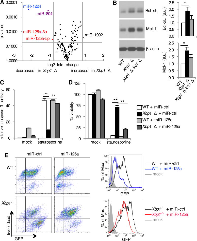
Fig. 6. IRE1α mediates reduction in pro-apoptotic miR-125a.
(A) BMDMs were cultured from Xbp1flox/flox ESR Cre+ (Xbp1 Δ), or Cre- littermate (WT) mice in the presence of tamoxifen. Volcano plot demonstrating distribution of microRNAs between WT and Xbp1 Δ BMDMs measured using the NanoString nCounter assay. Data are from one experiment with quadruplicates. (B) BMDMs were cultured from Xbp1flox/flox ESR Cre+ (Xbp1 Δ), Xbp1flox/flox Ern1flox/flox ESR Cre+ (Xbp1 Δ Ire1α Δ) or Cre- littermate (WT) mice in the presence of tamoxifen. The relative abundance of Bcl-xL, Mcl-1 and β-actin in the cell lysates was determined by Western blotting and densitometry. The ratio of Bcl-xL or Mcl-1 to β-actin is shown, normalized to WT. Data are means ± SD from three independent experiments. a.u., arbitrary units.(C and D) WT and Xbp1−/− MEFs were transfected with negative control microRNA mimetic (miR-ctrl) or miR-125a mimetic. Cells were left untreated (mock) or treated with staurosporine. Seven hours later, caspase-3 activity was assessed by measuring fluorometric substrate cleavage, and is shown relative to untreated WT cells (C). Twenty-four hours after treatment, viability was assessed by measuring MTS reduction (D). Data are means ± SD of three replicates and are representative of two experiments. (E) MicroRNA transfected MEFs were infected with VSV-GFP for 24 hours. Cell death was then assessed with a membrane impermeant, amine-reactive fluorescent dye, which was measured by flow cytometry. The extent of infection was determined by measuring the relative abundance of GFP by flow cytometry. Data are from one experiment representative of two independent experiments. *P < 0.01, **P < 0.001, unpaired t test.

