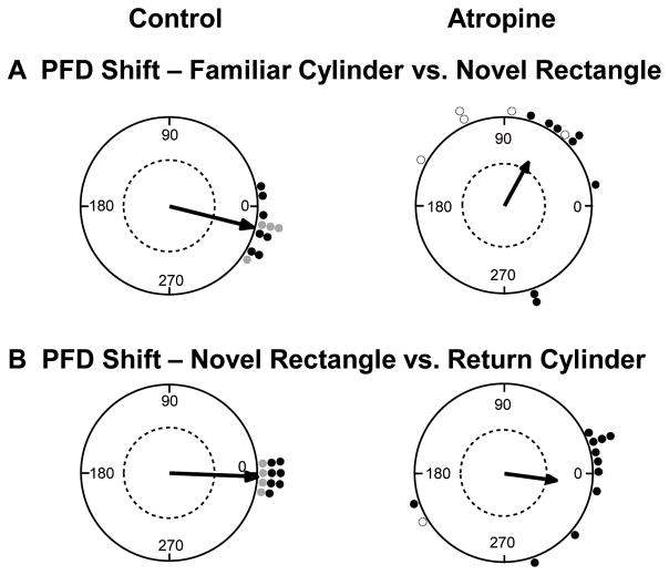
Figure 4.
Preferred firing direction (PFD) shifts in the dual chamber apparatus. A) Control HD cells (left) showed a relatively consistent preferred direction as the animal walked from the cylinder to the novel rectangle, suggesting the HD signal was maintained by path integration. In contrast, most HD cells in the atropine group (right) shifted toward the visual cue card (90° CCW), suggesting path integration failed to maintain the HD signal between arenas, and that the cue card in the rectangle may have driven the shift in the cells’ PFDs. B) Upon return to the familiar cylinder, control HD cells (left) realigned to the cue card in the cylinder. Atropine HD cells (right) also realigned to the cue card, albeit with less precision than the control group. Dashed line indicates .05 significance criterion. Open circles represent HD cells that shifted > 90° within the recording session. For the control group, black points represent HD cells recorded from the ADN (Taube & Burton, 1995) and gray points represent HD cells recorded from the lateral mammillary nuclei (Yoder et al., 2015).

