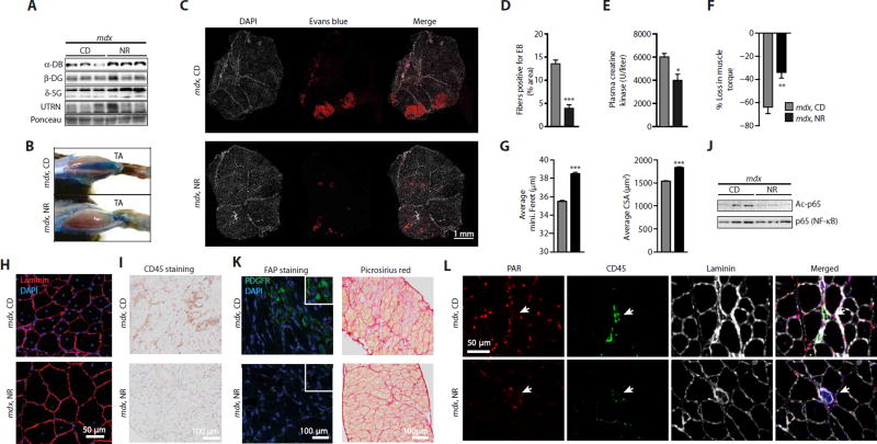
Fig. 5. NAD+ repletion causes compensatory increases in the expression of structural proteins and protects muscle from damage and fibrosis.
Sixteen-week-old mdx mice received a dietary supplement with NR (400 mg/kg per day) for 12 weeks. (A) NR-treated mdx mice exhibited increased α-dystrobrevin (α-DB) (CD, 1.00 ± 0.35; NR, 1.61 ± 0.09) and δ-sarcoglycan (δ-SG) (CD, 1.00 ± 0.15; NR, 2.24 ± 0.43; P = 0.026) protein expression compared to HSP90 (Fig. 4D) as a loading control in gastrocnemius extracts. NR attenuated mdx muscle damage as evidenced through (B) Evans Blue staining of both TA muscle and (C) sections of gastrocnemius and soleus muscles [red, Evans Blue (EB); white, 4′,6-diamidino-2-phenylindole (DAPI)], (D) quantified using ImageJ software (mdx, n = 6; mdx NR, n = 12). (E) NR also reduced basal plasma creatine kinase levels (mdx, n = 5; mdx NR, n = 6). (F) mdx mice treated with NR demonstrated an attenuation in the percent loss of torque in quadriceps, after muscle damage induced by lengthening contractions (mdx, n = 7; mdx NR, n = 6). (G) Increases in the average minimal Feret’s diameter (in micrometers) and cross-sectional area (CSA) (in square micrometers) of tibialis anterior muscle fibers, indicating increases in fiber size with NR treatment in mdx mice. These data were acquired from images stained with DAPI and laminin [(H) shows a representative image]. Quantification of images was performed with ImageJ software (8 weeks of NR treatment, n = 5). (I) Similarly, reduced CD45 staining was seen in diaphragms of mdx mice treated with NR. (J) Reduced acetylation of p65 (NF-κB) protein after NR in quadriceps (CD, 1.00 ± 0.15; NR, 0.38 ± 0.16; P = 0.023; n = 3). (K) FAPs expressing mesenchymal PDGFRα were decreased in diaphragms of mdx mice treated with NR (n = 3). Also, reductions in the fibrosis of mdx diaphragms were observed with less Picrosirius red staining in transverse muscle sections of NR-treated mice. (L) PARylation intensity in skeletal muscle nuclei of mdx mice is reduced with NR, as shown with immunohistological staining of tibialis anterior muscle with PAR, CD45, and laminin antibodies.

