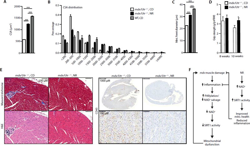
Fig. 6. mdx/Utr−/− mice recover skeletal muscle function and exhibit improved heart pathology with NR treatment.
Three-week-old mdx/Utr−/− mice received a dietary supplement with NR (400 mg/kg per day) for 5 to 7 weeks. Images stained with DAPI and laminin were used to quantify increases in the (A) average and (B) distribution of mdx/Utr−/− mouse muscle fiber cross-sectional areas (in µm2) of the tibialis anterior muscle (mdx/Utr−/−n = 3; 7 weeks of NR treatment). (C) NR-treated mdx/Utr−/− mice showed an increase in the minimal Feret’s diameter (in micrometers) (mdx/Utr−/−n = 3; 7 weeks of NR treatment). Quantification of images was performed with ImageJ software. (D) As evidence for the therapeutic effectiveness of NR treatment, mdx/Utr−/− mice grip strength was improved from 8 weeks (mdx/Utr−/−n = 4; mdx/Utr−/− NR, n = 7; 5 weeks of NR treatment) to 10 weeks of age (mdx/Utr−/−n = 3; mdx/Utr−/− NR, n = 5; 7 weeks of NR treatment). BW, body weight. (E) Sections of heart tissue were (immuno) histochemically stained with Masson’s trichrome, hematoxylin and eosin, and CD45, showing a reduction of cardiac fibrosis, necrosis, and macrophage infiltration, respectively, in the ventricles of mdx/Utr−/− mice at 4 weeks of age and treated with NR for 6 weeks (representative images from n = 3 mdx/Utr−/− and n = 3 mdx/Utr−/− NR). (F) Scheme summarizing the SIRT1-dependent effects of NR on mdx mouse muscle.

