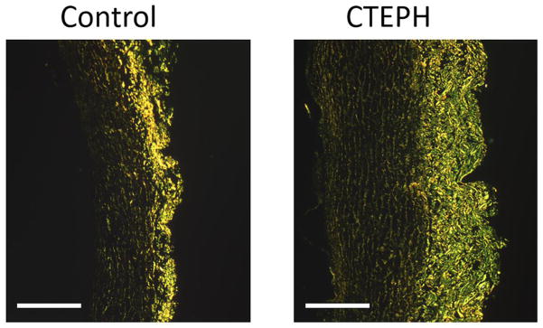
Fig. 4.
PA collagen fibers (bright yellow/green) imaged with picrosirius red staining viewed using polarized light for a control (left) and CTEPH (right) sample (scale bar = 500 μm). Collagen content and wall thickness were significantly increased in the CTEPH group, indicating arterial remodeling with chronic embolization. Percent collagen content was calculated by dividing the area marked positive for the collagen by the total tissue area.

