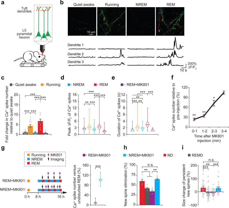
Figure 5. Dendritic Ca2+ spikes occurring during REM sleep are important for new spine elimination and strengthening.
(a) Two-photon Ca2+ imaging of apical tuft dendrites of L5 pyramidal neurons in head-restrained mice on a treadmill during quiet awake, running, NREM sleep and REM sleep. (b) Ca2+ imaging of apical tuft dendrites under various states. Ca2+ fluorescence traces of three dendrites over 1 min are shown. Images at 3 timepoints with a 10-s interval are represented by three different colors. Scale bar, 10 μm. (c,d) The number and peak amplitude of dendritic Ca2+ spikes during REM sleep were comparable to those during running but significantly larger than those during either NREM sleep, quiet awake state or REM sleep with MK801 application (for number: P = 1.82 × 10⁻⁵, 0.082, 9.37 × 10⁻⁵ and 1.66 × 10⁻⁴ for REM sleep vs. quiet awake, running, NREM sleep and REM sleep + MK801, respectively; n = 13, 12, 9, 13 and 8 quiet awake, running, NREM, REM and REM + MK801 mice, respectively; for peak amplitude: P = 2.82 × 10⁻⁴, 0.373, 9.09 × 10⁻⁸ and 1.77 × 10⁻¹² for REM sleep vs. quiet awake, running, NREM sleep and REM sleep + MK801, respectively; n = 89, 124, 88, 274 and 47 spikes for quiet awake, running, NREM, REM and REM + MK801, respectively). (e) The durations of dendritic Ca2+ spikes during REM sleep were significantly larger than those during other states (P = 3.01 × 10⁻¹⁷, 0.0007, 1.88 × 10⁻¹⁰ and 1.47 × 10⁻¹³ for REM sleep vs. quiet awake, running, NREM sleep and REM sleep + MK801, respectively). (f) Brief injection of MK801 (3 pulses, 50 ms each) into the primary motor cortex blocked dendritic Ca2+ spikes during quiet awake over the next 2–3 min (n = 10 mice). (g) More than 90% of dendritic Ca2+ spikes during REM sleep were blocked after pulsed injection of MK801 at the beginning of each REM sleep episode but not during NREM sleep (n = 46 episodes of REM sleep with MK801 injection from 4 mice; 51 episodes of NREM sleep with MK801 injection from 4 mice). (h) Injections of MK801 during REM sleep but not during NREM sleep, reduced the elimination rate of learning-induced new spines (ND vs. REM + MK801, P = 0.006; ND vs. NREM + MK801, P = 0.297; REM + MK801 vs. NREM + MK801, P = 0.006; n = 6, 5, 5 and 6 mice for ND, REMD, REM + MK801 and NREM + MK801, respectively). (i) Injection of MK801 during REM sleep, but not during NREM sleep, reduced the size increase of persistent new spines formed after treadmill training (ND vs. REM + MK801, P = 0.0019; ND vs. NREM + MK801, P = 0.801; REM + MK801 vs. NREM + MK801, P = 0.0005; n = 25, 40, 26 and 29 new spines from 6, 5, 5 and 6 mice for ND, REMD, REM + MK801 and NREM + MK80, respectively). Data are presented as mean ± s.e.m. In d, e and g, box and whisker plots show the means (central dot), medians (central line in the box), ranges between 25th and 75th percentiles (box) and minimum–maximum range (whiskers). Each point in c and h represents data from one animal. **P < 0.01, ***P < 0.001, n.s. = not significant.

