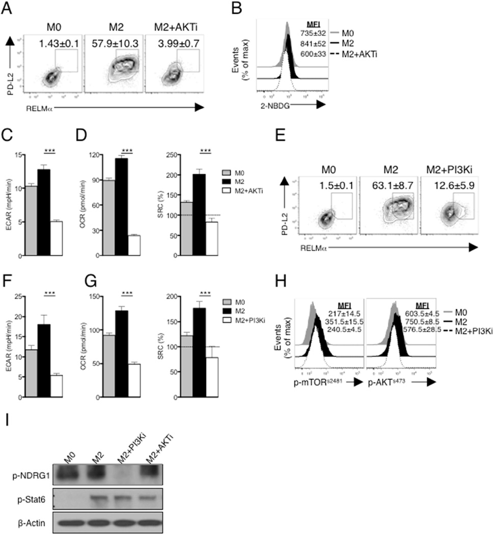
Figure 3. PI3K/AKT signaling is essential for M2 activation.
(A) PD-L2 and RELMα expression by macrophages cultured for 24 hr in the absence (M0) or presence of IL-4 (M2) plus or minus triciribine (AKTi). (B) 2-NBDG staining of macrophages treated as in panel A. (C,D) Basal ECAR, basal OCR and SRC of macrophages treated as in A. (E) Expression of PD-L2 and RELMα by M0 macrophages, or by M2 macrophages with or without LY294002 (PI3Ki) for 24 hr. (F,G) Basal ECAR, basal OCR and SRC in macrophages treated as in panel E. (H) Phosphorylation of mTORs2481 (p-mTORs2481) and AKTs473 (p- AKTs473) in macrophages treated as in panel E. (I) Phosphorylation of NDRG1 and Stat6 from unstimulated macrophages (M0) or macrophages stimulated with IL-4 (M2) for 3 hr in the presence or absence of PI3Ki and AKTi, assessed by immunoblot analysis. Data in A,B, E and H are from flow cytometry, and are from individual experiments, but numbers represent mean % or mean MFI, ± s.e.m, of data from three or more independent experiments. In C, D, F and G, data are mean ± s.e.m. of technical replicates from one experiment representative of three or more independent experiments. Data in I are from one experiment representative of three independent experiments. ***P < 0.0001.

