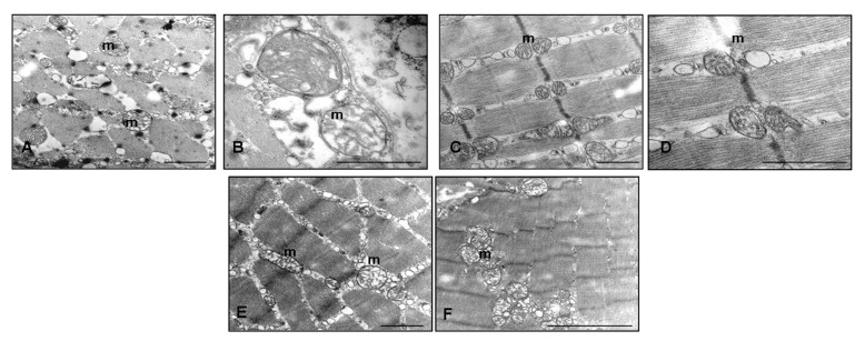
Figure 3.
Skeletal muscle ultrastructure. Transmission electron microscopy photomicrographs of: reserpine-induced myalgia rats (A,B); rats treated with reserpine and then with only tap water for two months (C,D); and rats treated with reserpine and then with melatonin at the dose of 5 mg/kg/day for two months (E,F). Scale bar: 1 μm. (m) identifies the mitochondria.

