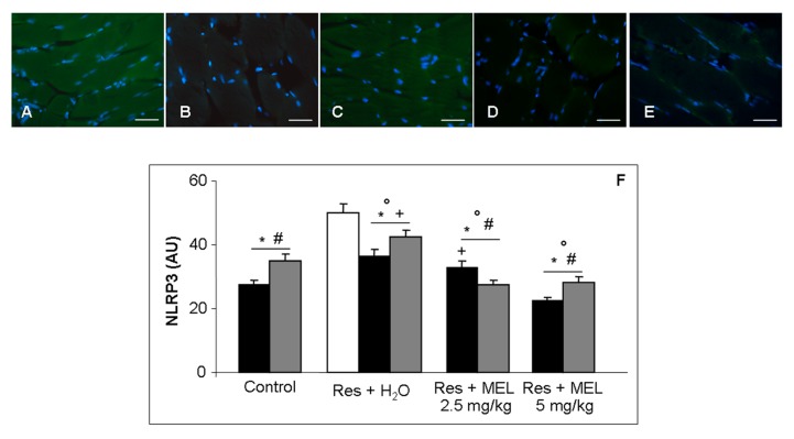
Figure 7.
Skeletal muscle inflammatory markers. Immunofluorescence photomicrographs of gastrocnemius muscle inflammosome NLRP3 expression of: reserpine-induced myalgia rats (A); controls (B); rats treated with reserpine for two months (C); rats treated with reserpine plus melatonin at the dose of 2.5 mg/kg/day for two months (D); and rats treated with reserpine and then with melatonin at the dose of 5 mg/kg/day for two months (E). Nuclei were stained with DAPI (blue). Scale Bar: 20 µm. The graph summarizes the histomorphometrical analyses, expressed in arbitrary units (AU), of inflammosomeNLRP3. ANOVA, two-way analysis of variance; * p ≤ 0.05 vs. Reserpine four days; # p ≤ 0.05 vs. Reserpine plus tap water; + p ≤ 0.05 vs. Reserpine plus melatonin 5 mg/kg/day for 2 months and ° p ≤ 0.05 vs. Controls. H2O: tap water; MEL: melatonin; Res: reserpine.

