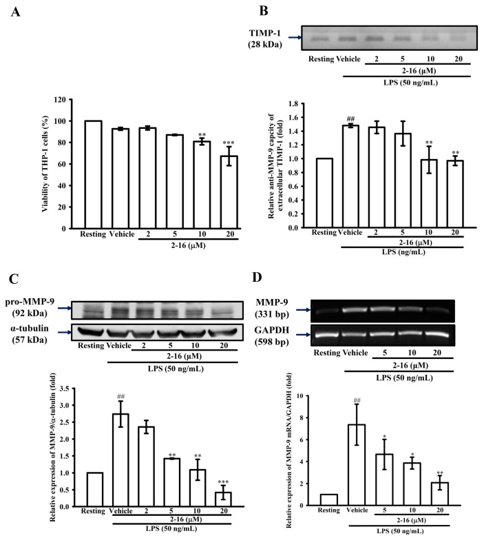
Figure 3.
WK2-16 inhibits MMP-9 protein and mRNA expression induced by LPS in THP-1 cells without cellular toxicity. (A) THP-1 cells (5 × 10⁵ cells/0.5 mL) were dispensed onto 24-well plates and were treated with the indicated concentrations of WK2-16 (2, 5, 10 and 20 μM) or vehicle for 24 h. Cell viability was quantified by the ability of mitochondria to reduce the tetrazolium dye 3-(4,5-dimethylthiazol-2-yl)-2,5-diphenyl tetrazolium bromide (MTT) in viable cells. (B) The activity of tissue inhibitor of matrix metalloproteinase-1 (TIMP-1) as assessed by reverse zymography of conditioned media from THP-1 cells treated with the indicated concentrations of WK2-16 (2, 5, 10 and 20 μM) or vehicle or 15 min before exposure to LPS (50 ng/mL) for 24 h. (C,D) THP-1 cells (106 cells/mL) were dispensed onto six-well plates and were treated with LPS (50 ng/mL) for: 24 h (C); or 8 h (D) at the indicated concentrations of WK2-16 (2, 5, 10 and 20 μM) or vehicle for 15 min before treatment with LPS. Cell lysates were obtained and analyzed for MMP-9 protein expression by Western blotting or for MMP-9 mRNA expression by RT-PCR. The data are represented as the means ± S.D. from three independent experiments. ## p < 0.01 compared with the resting condition, * p < 0.05, ** p < 0.01 and *** p < 0.001 compared with the vehicle.

