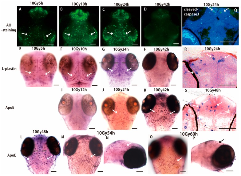
Figure 1.
Sequential process of microglial activation following γ-ray irradiation. Acridine orange (AO)-positive apoptotic neurons started to form rosette-shaped clusters 5 h after the irradiation of γ ray (10 Gy) (A). Number of clusters increased and they were located in the marginal area of the optic tectum (OT) 10–24 h after the irradiation (B,C) then disappeared 42 h after the irradiation (D). The distribution of l-plastin mRNA revealed by Whole-Mount In Situ Hybridization (WISH) was identical to that of AO-positive apoptotic neurons during 5–42 h after the irradiation (E–H). In a histological section of the WISH-processed brain 24 h after the irradiation (G) prepared at the dotted line, l-plastin expressing microglia were distributed in the marginal area of the irradiated OT (arrows in R) in the same manner as that of cleaved-caspase 3 positive apoptotic neurons (arrows in Q). By contrast, no ApolipoproteinE (ApoE)-expressing microglia were present 12 h after the irradiation (I). ApoE-expressing microglia started to appear 24 h after the irradiation (arrow in J) and they increased throughout the whole brain 42–48 h after the irradiation (arrows in K,L). A histological section of the WISH-processed brain 48 h after irradiation (L) prepared at the dotted line in L shows that the activated microglia changed their cell appearance from ramified to amoeboid morphology (arrows in S). Number of ApoE-expressing microglia decreased 54 h after the irradiation (arrows in M,N) and completely disappeared within the brain, but accumulated on the dorsal surface of the brain 60 h after the irradiation (arrow in O,P). AO-stained and WISH-processed brains in A–M and O show dorsal views, and (N,P) show lateral views. Scale bars = 100 μm.

