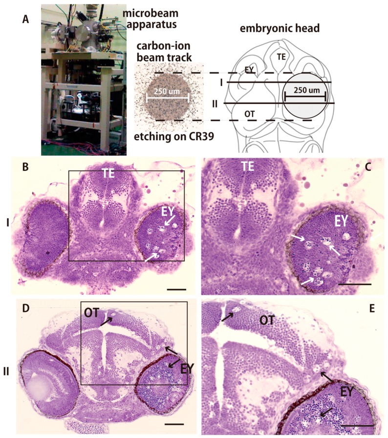
Figure 2.
Apoptotic neuronal deaths were induced only in the targeted irradiated area 24 h after irradiation. (A) A collimated carbon ion microbeam (250 μm diameter) characterized by etching on CR39, was used to locally irradiate the right hemisphere of optic tectum (OT). Nissl-stained frontal sections of the locally irradiated brain were prepared at the solid lines (I and II in A) and shown in B–E, respectively. Many pyknotic cells were induced only in the right retina (arrows in B–E) and the right hemisphere of the OT (arrows in D,E). (B,C) show the frontal sections prepared at the solid line labeled I in A, and (D,E) show the frontal sections prepared at the solid line labeled II in A. OT, optic tectum; TE, telencephalon; EY, eye. Scale bars = 50 μm.

