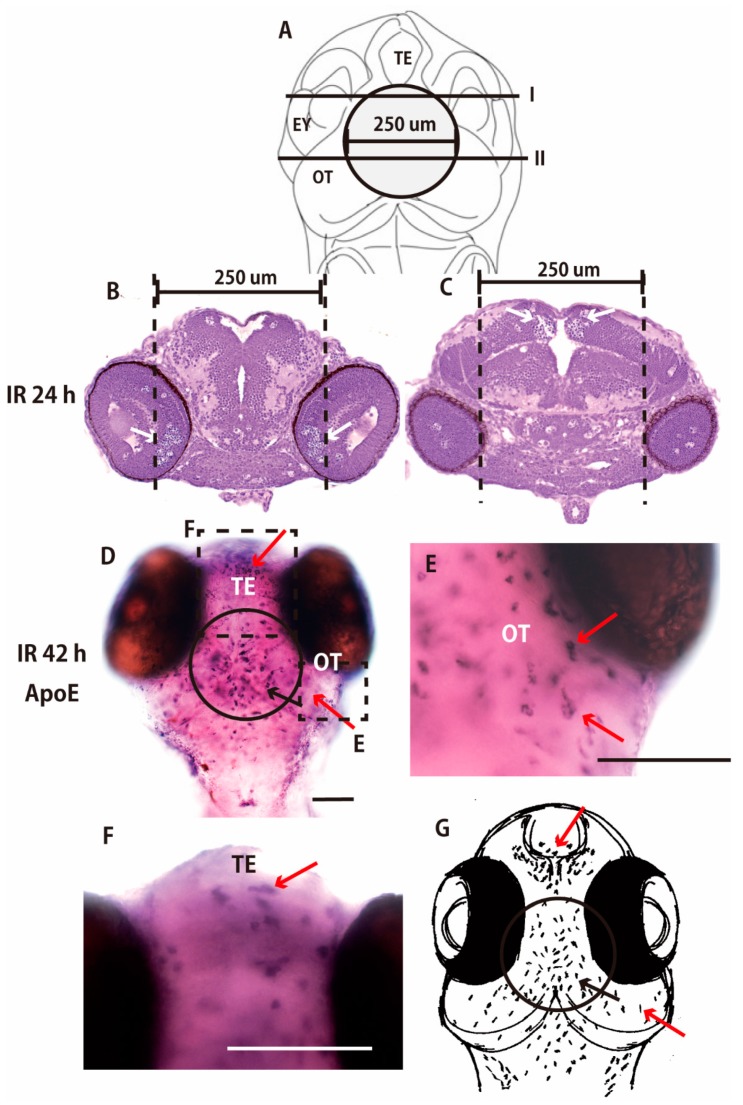
Figure 4.
Irradiation targeted to the center of OT of medaka embryonic brain. Nissl-stained frontal sections of the brain 24 h after the targeted irradiation were prepared at the solid lines of I and II in (A) and shown in (B,C), respectively. Many pyknotic cells were induced in the limited area of the retina (arrows in B) and the central part of OT (arrows in C) where microbeam irradiation was targeted (dotted lines in B,C). ApoE-expressing activated microglia at the late phase of phagocytosis were present 42 h after the irradiation, not only in the irradiated area of the central part of OT (circled area in D), but also beyond the irradiated area of OT (red arrows in E) and in the telencephalon (red arrow in F). A schematic drawing of the abscopal effect of microglial activation is shown in G (red arrows in G). Scale bars = 100 μm.

