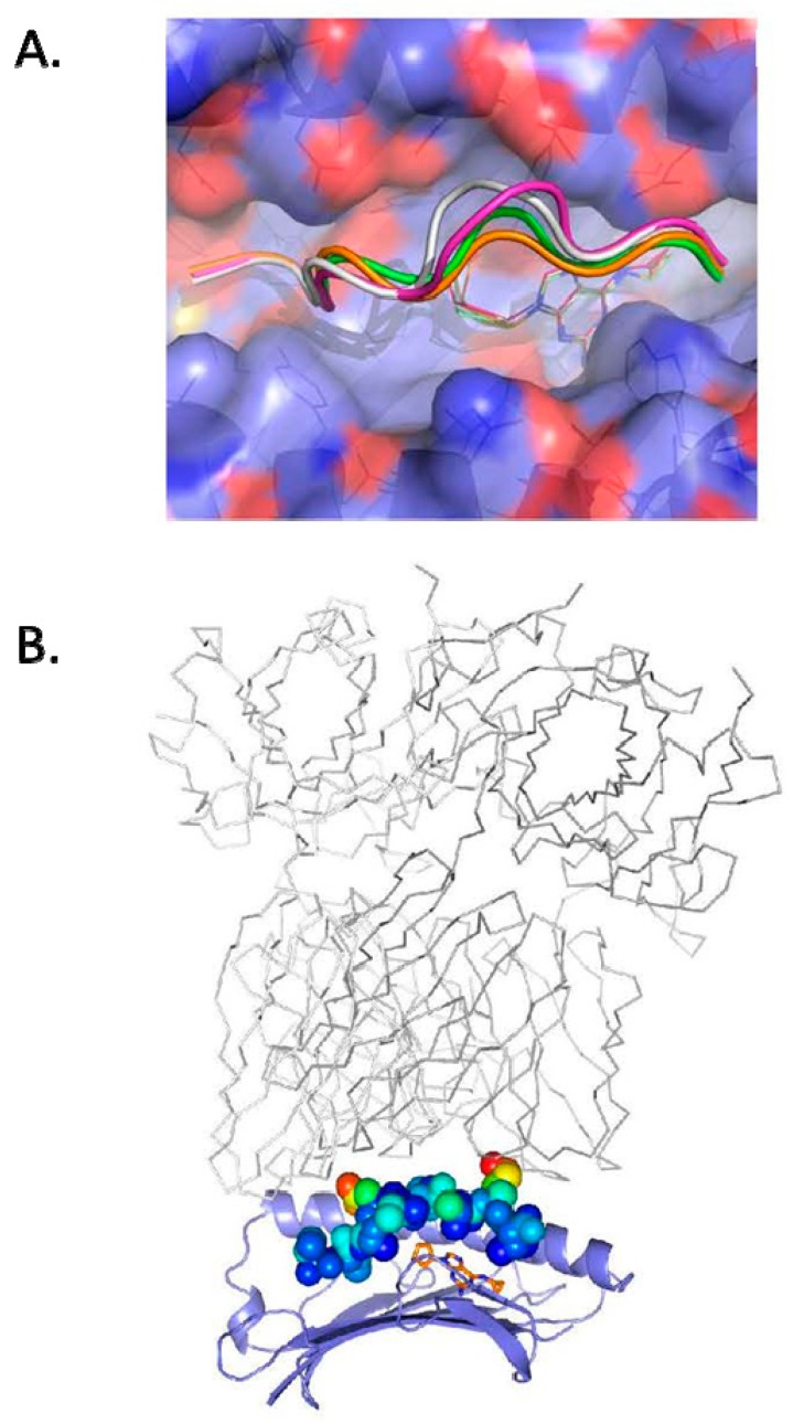
Figure 1.
The crystal structure of self peptide VTTDIQVKV SPT5a 976–984 complexed to abacavir and HLA-B*57:01 reveals solvent accessible side chains available for recognition by T cells. (A): abacavir is shown in the crystal structure of VTTDIQVKV SPT5a 976–984/HLA-B*57:01 as gold sticks. Binding of abacavir was similar to solved structures of other peptides complexed with abacavir and HLA-B*57:01: PDB code 5U98, gold for carbon, 3VRI, magenta, 3VRJ, white, 3UPR, green. The molecular surface of HLA-B*57:01 is shown as violet for carbon, blue for nitrogen, red for oxygen. (B): the crystal structure of VTTDIQVKV SPT5a 976–984 complexed to abacavir and HLA-B*57:01 is shown with the peptide as spheres colored by B factor. P4D and P8K exhibiting the greatest degree of flexibility and solvent exposure. The HLA-B restricted TCR (gray lines) from 2NX5 was superimposed on HLA-B*57:01 to show a conventional binding mode of TCR with respect to peptide and HLA.

