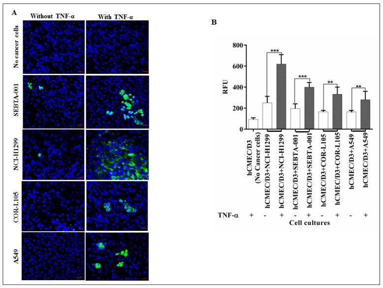
Figure 2.
The role of CD62E in adhesion of NSCLC cells to brain endothelium: (A) Qualitative adhesion of NSCLC cells onto brain endothelium monolayer. Green fluorescently tagged NSCLC cells were applied onto the hCMEC/D3 monolayer and incubated for 90 min with and without activation via TNF-α. Non-adherent cells were washed and co-cultures were fixed and examined by confocal microscopy; (B) quantitative adhesion of NSCLC cells. hCMEC/D3 cells were seeded into 96-well plate followed by seeding of green fluorescently tagged NSCLC cells on the hCMEC/D3 monolayer and incubated for 90 min incubation. Non-adherent cells were washed out and adherent cells were lysed followed by quantification via a microplate reader at 480–520 nm. Results showed a strong decrease in adhesion caused by absence of TNF-α (White bar) compared to TNF-α stimuli. n = 3, *** p < 0.001 and ** p < 0.01. Scale bar = 20 µm. Data is expressed as ±SE.

