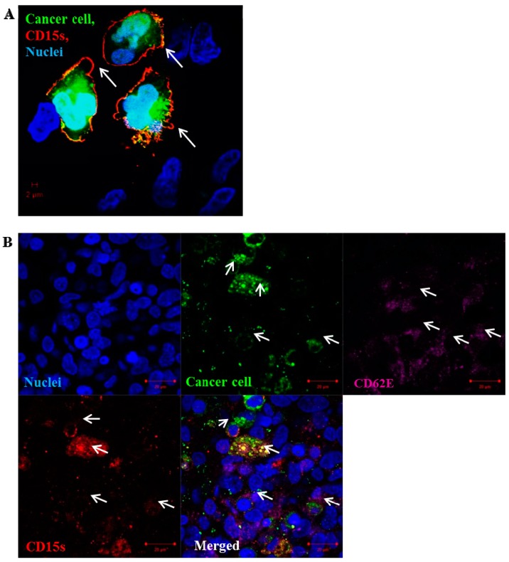
Figure 5.
Localisation of CD15s on the surface of adherent SEBTA-001 cells at the site of adhesion. Confocal image of green fluorescently-labelled adherent brain to lung metastatic cancer cells (SEBTA-001) on a monolayer of activated hCMEC/D3 cells (blue). (A) Expression of CD15s (red) (white arrows) was seen on the edges of adherent cells SEBTA-001 (green) at the site of adhesion on brain endothelial cells; (B) expression of CD15s (red) (white arrows) was observed on surface of adherent cancer cells SEBTA-001 (green) and CD62E (purple) (white arrows) was seen on the monolayer of activated brain endothelial cells (hCMEC/D3) at adhesion site of cancer cell–brain endothelium. Scale bar = 20 µm.

