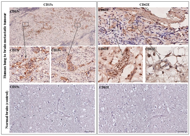
Figure 6.
Representative immunohistochemistry images from one patient with lung–brain metastasis. (Left panel) CD15s was detected in tumour core and infiltrated into non-neoplastic host brain tissue. No expression was seen in a normal brain section. (Right panel) CD62E was expressed on the inner lining of brain endothelial cells with no expression seen in normal brain sections. Images were obtained using an Ariol microscope (Leica, Wetzlar, Germany) at 20× and 40× magnification. Scale bar = 20 µm. n = 1.

