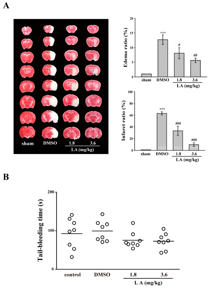
Figure 5.
Influence of LA on middle cerebral artery occlusion (MCAO)/reperfusion-induced brain injury and tail bleeding time in mice. Mice (male, 5–6 weeks old) were intraperitoneally administrated with DMSO (solvent control) or LA (1.8 and 3.6 mg/kg) for 30 min. (A) Mice were subjected to MCAO for 30 min followed by 24-h reperfusion. Immediately after sacrifice, coronal sections were cut and stained using 2,3,5-triphenyltetrazolium chloride; white areas indicate infarction, and red areas indicate normal tissues (left panel). Edema and infarct ratios (right panel) were calculated through image analysis and are reported as a ratio of the non-ischemic hemisphere. Infarct ratio was corrected for edema. Data are presented as the mean ± SEM. (n = 8). *** p < 0.001, compared with the sham-operated group; # p < 0.05, ## p < 0.01 and ### p < 0.001, compared with the DMSO group; (B) Bleeding was induced by severing the tail at 3 mm from the tail tip, and the bleeding tail stump was immersed in saline. Subsequently, the bleeding time was continually recorded until no sign of bleeding was observed for at least 10 s. Each point in the scatter plots graph represents a mouse (n = 8). The bars represent the median bleeding time of each group.

