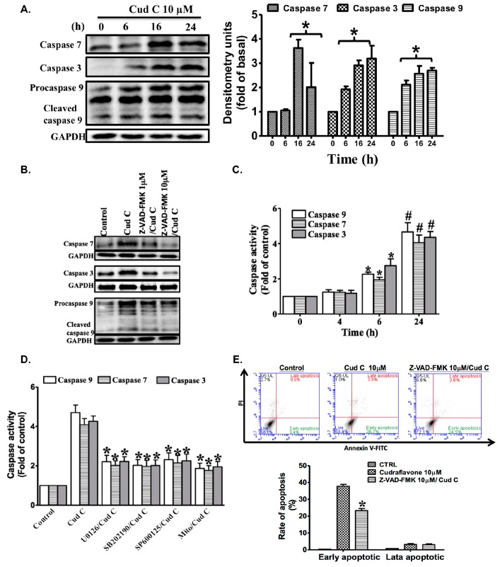
Figure 5.
(A) Effects of cudraflavone C on the expression status of caspase-7, caspase-3, and caspase-9 in A375.S2 cells over various time periods (0–24 h), as determined by Western blotting; (B) In addition, cells were pretreated for 1 h with the caspase inhibitor Z-VAD-FMK and then treated with cudraflavone C for 16 h, and the levels of active caspase-7, -3, and -9 were evaluated by Western blotting; (C,D) Cells were treated with cudraflavone C for indicated times. The caspase activity was analyzed by using caspase-3, -7, and -9 colorimetric assay kits; (E) Confluent cells were pre-incubated with or without inhibitors of ERK1/2 (U0126), p38 (SB202190), JNK1/2 (SP600125) and MitoTEMPOL (mitochondria-targeted antioxidant). After incubation for 1 h, cells were treated with cudraflavone C (10 µM) for 24 h. The activities of caspases were analyzed by using caspase-3, -7, and -9 colorimetric assay kits. Results are representative of three independent experiments. * p < 0.05, # p < 0.01 compared to the control group.

