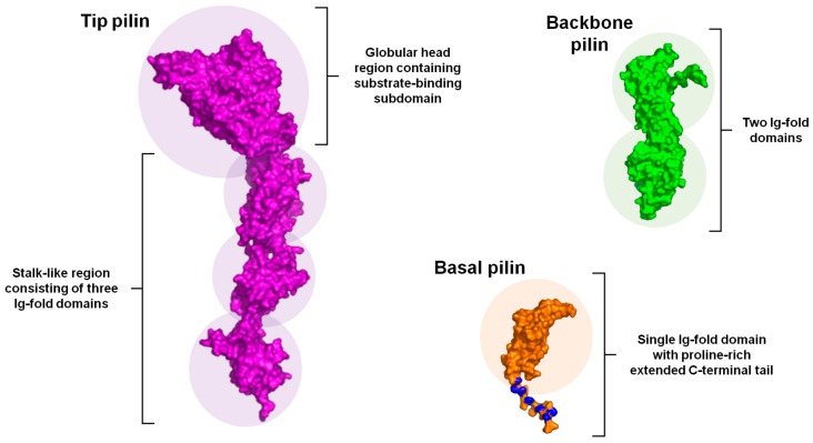
Figure 2.
Three-dimensional view of the tip, backbone, and basal pilin structures within a typical sortase-dependent pilus. Shown are representative tertiary structures of the three types of pilin subunits. The structure of the tip pilin is comprised of a stalk-like region consisting of three Ig-fold domains (e.g., CnaA or CnaB), upon which rests a globular head region that contains the substrate-binding domain. The C-terminal LPXTG peptide is normally situated within the bottom subdomain. The backbone pilin structure is shown having two Ig-fold domains, with the linking lysine and the LPXTG peptide located within the upper and lower domains, respectively. The basal pilin structure is depicted with a single Ig-fold domain that includes a proline-rich (blue) extended C-terminal tail. All domain regions are highlighted by circular background shading. Structural visualization of the tip, backbone, and basal pilins was rendered by PyMol (http://www.pymol.org/) using protein atomic coordinates (2WW8, 3B2M, and 3LKQ, respectively) retrieved from the RCSB Protein Data Bank (http:// http://www.rcsb.org/pdb/home/home.do).

