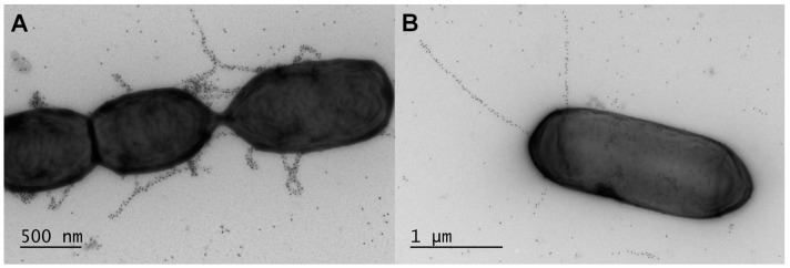
Figure 3.
Visualization of native sortase-dependent piliation in lactobacilli by immuno-electron microscopy. Immunogold pilin-protein labeling and electron microscopy (EM) of bacterial cells were done using established techniques [89]. Long protruding structures representing the SpaCBA pili in L. rhamnosus GG (A) and LrpCBA pili in L. ruminis ATCC 25644 (B) are identified by immuno-labeling first with polyclonal antibody specific for the backbone-pilin subunit (SpaA and LrpA, respectively) and then protein A-conjugated gold particles (10 nm in diameter; black dots), followed by negative staining and electron microscopy (EM). Scale bars for each electron micrograph image are found at the bottom-left corner.

