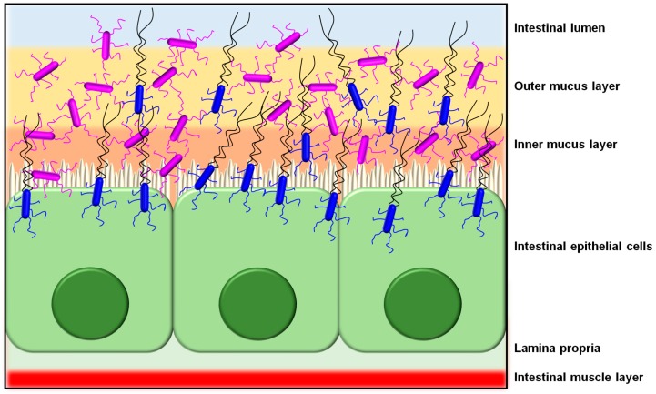
Figure 4.
Schematic depiction showing the presumptive binding of piliated lactobacilli to the intestinal mucosa epithelium. Based on substrate-binding capacities, L. rhamnosus cells (purple) with their mucoadhesive SpaCBA pili would be predominantly found within the outer and inner mucus layers, whereas flagellated L. ruminis cells (blue) would be able to maneuver through this mucosal barrier and then, via their ECM-binding LrpCBA pili, attach themselves to the single cell layer of intestinal epithelial tissue. This cartoon image is representative only, and includes various aspects and features that are not drawn to scale.

