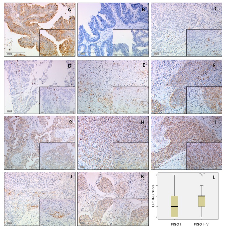
Figure 1.
All images are at 10× magnification with an insert at 25× magnification. (A) Positive control of ovarian cancer metastasis in the colon shows cystoplasmatic and membrane-associated staining; (B) Negative control of ovarian cancer metastasis in the colon; (C) Squamous cell carcinoma Immunoreactive score (IRS) 1; (D) Adenocarcinoma IRS 1; (E) Squamous cell carcinoma IRS 4; (F) Adenocarcinoma IRS 4; (G) Adenocarcinoma carcinoma IRS 8; (H) Squamous cell carcinoma IRS 8, (I) Squamous cell carcinoma IRS 9; (J) Prostaglandin E receptor type 3 (EP3) staining of an Fédération Internationale de Gynécologie et d’Obstétrique (FIGO) Ib diagnosed IRS 2 stained squamous cell carcinoma; (K) EP3 staining of an FIGO 4 diagnosed IRS 4 stained squamous cell carcinoma; (L) Boxplot of FIGO I and FIGO II–IV cases with median IRS.

