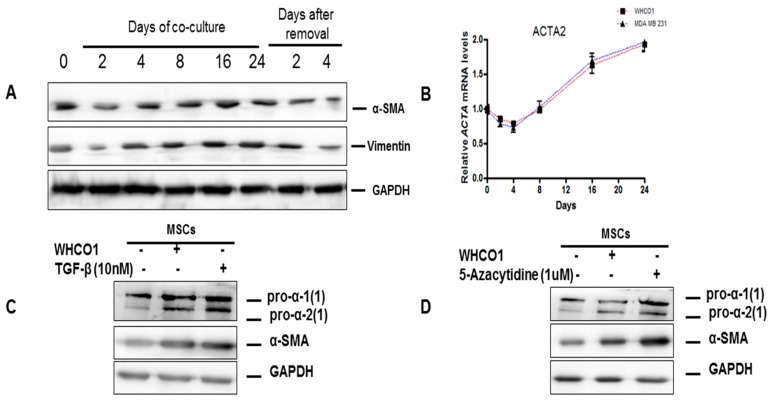
Figure 3.
Cancer cells trigger MSCs differentiation into tumor associated fibroblasts via the transforming growth factor-β (TGF-β) /Smad signaling pathway. For the co-culture experiments cells were co-cultured in 6-transwell plates (size of pore: 0.4 μm, Polycarbonate membrane, Costar, Corning, Cambridge, MA, USA). Mesenchymal stem cells (5 × 10⁵ cells) were cultured in the upper insert and cancer cells (WHCO1 and MDA MB 231) (5 × 10⁵ cells) were cultured in the lower compartment. Empty inserts were used for the control group (no cells) and a mixture of MSCs medium and cancer cell medium (1:1) was used. Medium was changed every 3 days for longer incubation periods and fresh TGF-β and reagents were added. TGF-β and all reagents were added to the media to the final concentrations as shown. At specific time points or at the end of the experiment, cells (cancer cells and MSCs) were harvested and used in various analyses. (A) Western blot analysis of lysates from MSCs co-cultured with WHCO1 cells for 24 days showing α-smooth muscle actin (α-SMA) and vimentin protein levels. Glyceraldehyde 3-phosphate dehydrogenase (GAPDH) was used as a loading control. (B) Real time quantitative reverse transcription polymerase chain reaction (RT-qPCR) analysis was performed to assess the expression of Actin, alpha2, smooth muscle, aorta (ACTA2) (α-SMA gene) in MSCs co-cultured with WHCO1 and MDA MB 231 cancer cells over a 24 day period; (C,D) western blot analysis of lysates from MSCs co-cultured with WHCO1 cells for 16 days or after the addition of 10 nM TGF-β (C) or 1 μM 5-azacytidine (D) for 48 h showing the expression of type I collagen and α-SMA; (E,F) western blot analysis of lysates from MSCs co-cultured with MDA MB 231 cells for 16 days or after the addition of 10 nM TGF-β (E) or the addition of 1 μM 5-azacytidine (F) for 48 h showing the expression of type I collagen and α-SMA.


