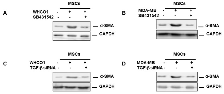
Figure 4.
WHCO1, MDA MB 231 cells and MSCs secrete TGF-β. Mesenchymal stem cells (5 × 10⁵ cells) were cultured in the upper insert and cancer cells (WHCO1 and MDA MB 231) (5 × 10⁵ cells) were cultured in the lower compartment as described in Figure 3. At specific time points or at the end of the experiment, cells (cancer cells and MSCs) were harvested and used in various analyses. (A,B) TGF-β inhibitor SB431542 was added to the co-culture media to a final concentration of 10 µM. Co-culture was continued for 16 days after which α-SMA protein levels was determined by western blot analysis. Glyceraldehyde 3-phosphate dehydrogenase (GAPDH) was used as a loading control. (C,D) MSCs were treated with TGF-β siRNA to a final concentration of 100 nM and co-culture was continued for 16 days. To maintain knockdown of TGF-β, subsequent transfections were done every other three days till the end of the experiment. Western blot analysis was performed to evaluate the α-SMA protein levels in MSCs lysates; (E,F) WHCO1 and MDA MB 231 cells were treated with TGF-β siRNA to a final concentration of 100 nM and co-culture was continued for 16 days. To maintain knockdown of TGF-β, subsequent transfections were done every other three days till the end of the experiment. Western blot analysis was performed to evaluate the α-SMA protein levels in MSCs lysates.


