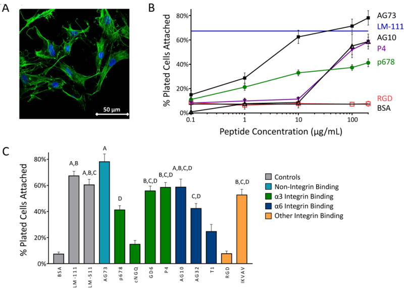
Figure 1.

(A) Primary human NP cells display consistent morphology and higher attachment on LM-111-coated tissue culture plastic (scale bar = 50 μm). (B) NP cells attach to peptide-coated surfaces at levels equivalent to that for LM-111, but only at higher peptide densities and for select peptides as shown here. (C) Cell numbers attaching to peptide coated surfaces for all screened peptides at 200 μg/mL. Conditions labeled with the same letter (A, B, etc) are equivalent and significantly different than the BSA control (p<0.05, ANOVA with post-hoc tests). SEM are shown.
