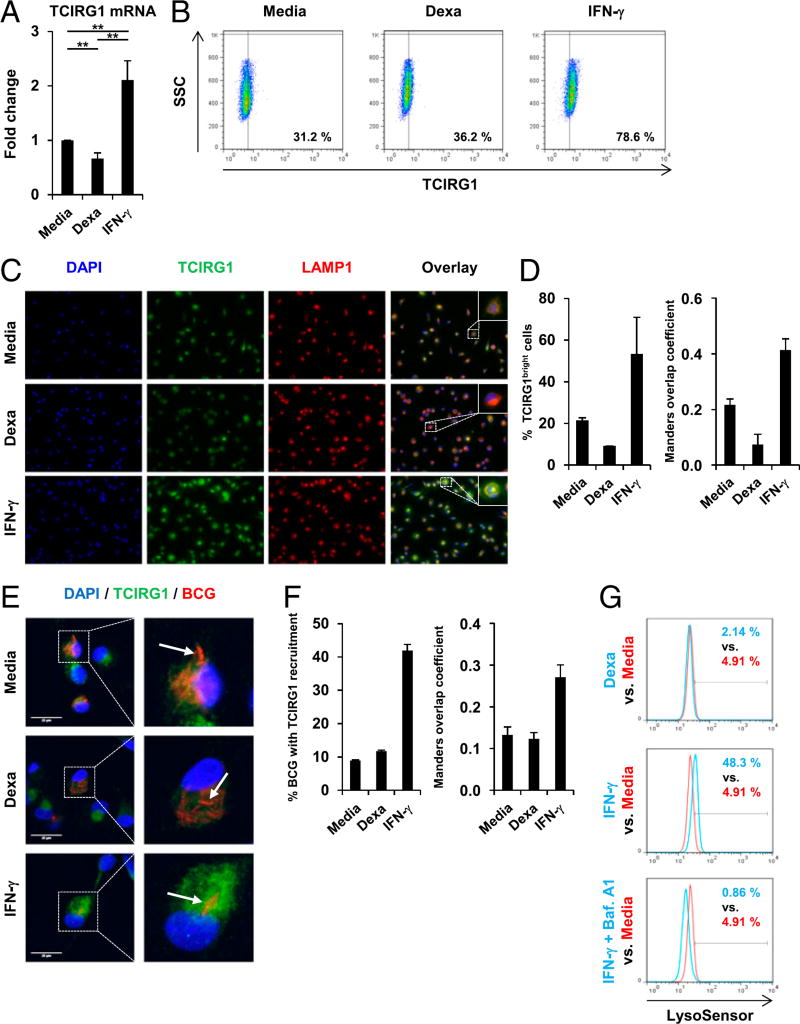
FIGURE 7.
IFN-γ triggers but dexamethasone (Dexa) fails to induce TCIRG1 expression, phagolysosome recruitment, and lysosome acidification in human macrophages. (A–C) MDMs were stimulated with Dexa (100 nM), IFN-γ, or media alone for 20 h. (A) Gene expression of TCIRG1 was assessed by quantitative PCR (mean fold change ± SEM, n = 12). (B) Intracellular TCIRG1 protein expression was determined by FACS analyses (% TCIRG1+ cells). (C) MDMs were fixed and immunostained with anti-TCIRG1 and anti-LAMP1. Original magnification ×40. (D) Quantification of (C). Left panel, Percentage of TCIRG1bright cells. Right panel, Manders overlap coefficient M2 (fraction of LAMP1 overlapping TCIRG1). (E) MDMs were infected with BCG (pMV261::dsRed) for 3 h and stimulated with Dexa (100 nM), IFN-γ, or media alone for 20 h, fixed, immunostained with anti-TCIRG1, and analyzed by confocal microscopy. Image brightness was enhanced equally across the entire image per journal policy. (F) Quantification of (E). Left panel, Percentage of BCG/BCG clusters with TCIRG1 recruitment. Right panel, Manders overlap coefficient M2 (fraction of BCG overlapping TCIRG1). (G) MDMs were stimulated with Dexa (100 nM), IFN-γ, IFN-γ and Bafilomycin A1 in combination, or media alone for 20 h and stained with LysoSensor Green. Acidification was determined by FACS analyses (% LysoSensor-positive cells). **p < 0.01.

