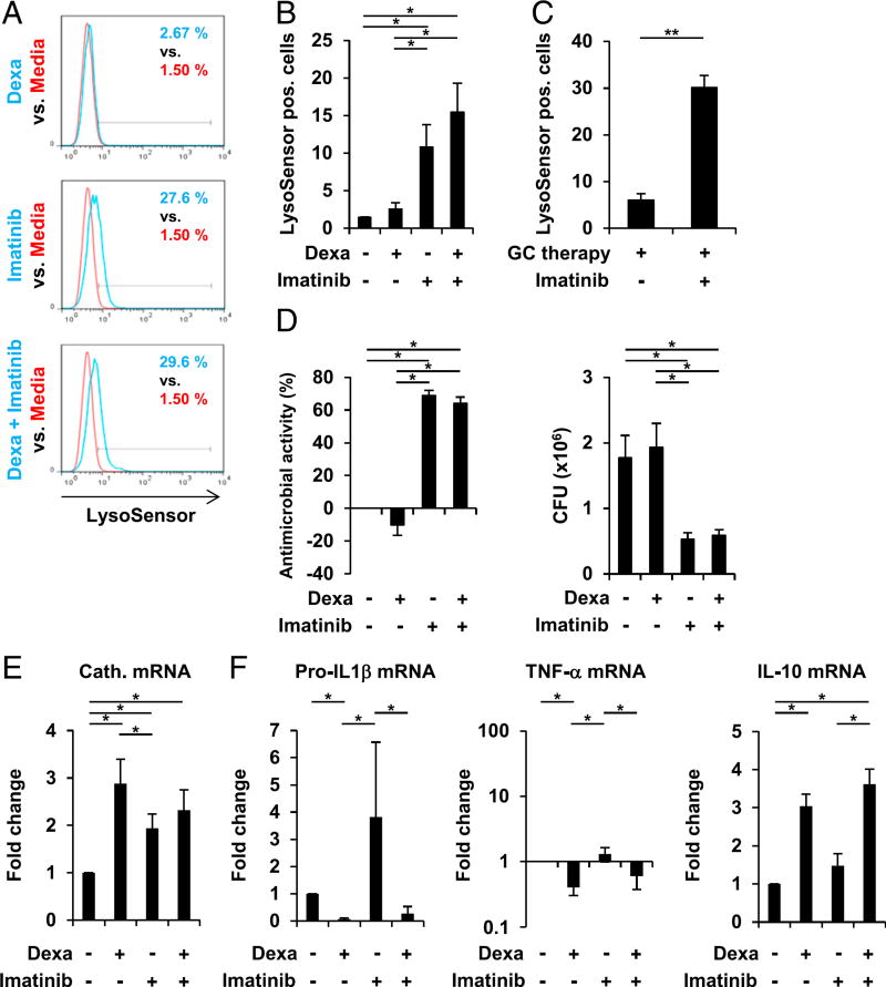
FIGURE 8.
Imatinib promotes lysosome acidification and antimycobacterial activity in dexamethasone (Dexa)-treated human macrophages. (A and B) MDMs were stimulated with Dexa (100 nM), Imatinib, Dexa and imatinib in combination, or media alone for 20 h. Cells were stained with LysoSensor Green. Acidification was determined by FACS analyses (% LysoSensor-positive cells). (A) Histogram plots from one representative donor out of seven. (B) Summary of seven donors (% LysoSensor-positive cells ± SEM, n = 7). (C) Monocytes from patients on long-term glucocorticoid therapy (GC therapy) were stimulated with imatinib or media alone for 20 h. Cells were stained with LysoSensor Green. Acidification was determined by FACS analyses (% LysoSensor-positive cells ± SEM, n = 4). (D) MDMs were infected with BCG for 3–4 h before stimulation with Dexa (100 nM), Imatinib, Dexa and imatinib in combination, or media alone for 3 d. Viable bacteria were quantified by CFU assay on day 3. Percentage antimicrobial activity relative to media (left panel) and CFU counts (right panel) are shown (± SEM, n = 5). (E) MDMs were stimulated as in (A). Cathelicidin mRNA expression was assessed by quantitative PCR (mean fold change ± SEM, n = 6). (F) MDMs were stimulated as in (A). Pro–IL1-β (n = 5), TNF-α (n = 5), and IL-10 (n = 5) mRNA expression were assessed by quantitative PCR (mean fold change ± SEM). *p < 0.05, **p < 0.01.

