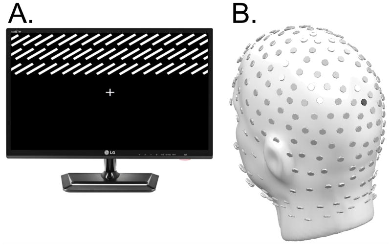
Figure 1.
Example Stimulus and EEG sensor array: (A) Representation of one of four gradient field stimuli (individual lines are enlarged for demonstration purposes, and thus there are fewer lines than were presented to participants). Gradient fields comprised lines oriented either at -75° or +75° relative to a vertical axis, and were located in either the upper or lower visual field relative to the fixation cross. The example illustrates a stimulus with a +75° orientation located in the upper visual field. (B) Layout of the 256-channel HydroCel Geodesic Sensor Array used for data acquisition in the current study. For reference, sensor POz is highlighted in black.

