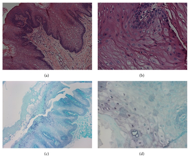
Figure 2.
Histological characteristics of OHL and in situ hybridization reaction. (a) Histopathological section, stained with H&E, showing stratified squamous epithelium, acanthosis, epithelial hyperplasia, cells resembling koilocytes, cells with opaque nucleus, and perinuclear halo (100x magnification). (b) Section stained with H&E showing cells resembling koilocytes (400x magnification). (c) In situ hybridization reaction with positivity for EBV (100x magnification). (d) In situ hybridization reaction with positivity for EBV (400x magnification).

