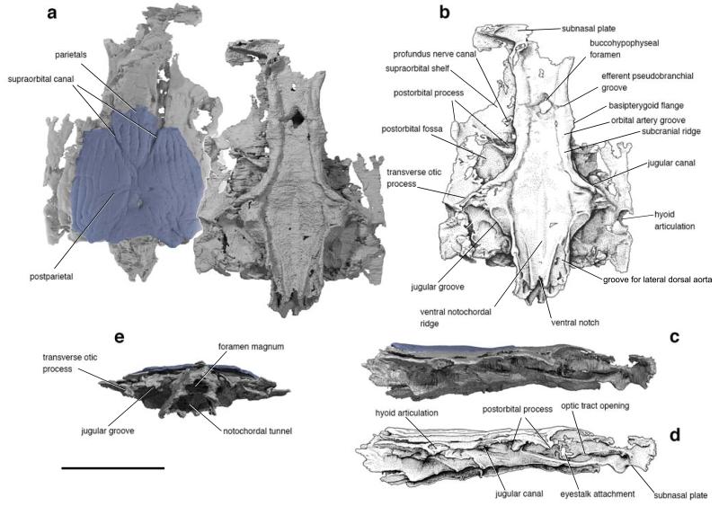
Figure 1. The skull of Janusiscus schultzei gen. et sp. nov. based on high-resolution computed tomography of GIT 496-6 (Pi.1384).
a, Dorsal (left) and ventral views. b, Interpretive drawing of ventral view. c, Right lateral view. d, Interpretive drawing of right lateral view. e, Posterior view. Scale bar, 5 mm.

