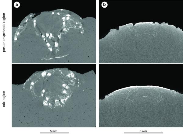
Extended Data Figure 4. Janusiscus lacks endochondral ossification.
a, The actinopterygian Kentuckia deani MCZ 5226, tomographs showing extensive and well-developed endochondral ossification in both the sphenoid (top) and otic (bottom) regions. Bright white objects in both panels are voids within spongy endochodral bone that have been diagenetically infilled with dense (likely iron) minerals. b, Janusiscus schultzei n. gen et sp. nov. GIT 496-6 [Pi.1384], tomographs showing lack of obvious endochondral ossification in either the sphenoid (top) or otic (bottom) regions. There is also no visual indication of endochondral bone in a break across the ethmoid region of this same specimen.

