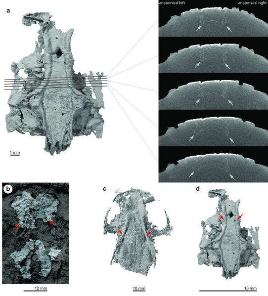
Extended Data Figure 5. Subcranial ridges in Janusiscus and early crown gnathostomes.
a, Reconstructed tomographs showing that the thickenings along the lateral margins of the sphenoid region of Janusiscus do not represent artefacts of post-mortem compression. b, The ‘acanthodian’ Ptomacanthus anglicus NHMUK PV P 24919a, silicone peel of specimen preserved in negative, dusted with ammonium chloride. Portions of the skull other than the neurocranium partially masked for clarity. c, The chondrichthyan Doliodus problematicus NBMG 10127/1a, reconstruction of neurocranium based on CT data. d, Janusiscus schultzei n. gen et sp. nov. GIT 496-6 [Pi.1384], reconstruction of neurocranium based on CT data. Red arrows in each panel indicate subcranial ridges.

