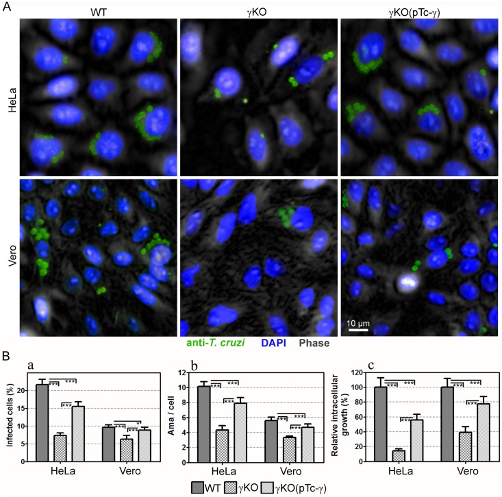
Fig 7. Effect of Trypanosoma cruzi AP-1 γ subunit (TcAP1-γ) gene knockout on T. cruzi γKO infection in mammalian cells.
Vero and HeLa cells were infected with WT, TcγKO (γKO) and TcγKO(pTc-γ) (complemented TcγKO) metacyclic trypomastigotes for 72 h. Then, they were labeled with the anti-T. cruzi serum (anti-TEMA) (detected using anti-rabbit IgG conjugated to Alexa Fluor 488) and DAPI (for DNA detection). Cells were imaged using the Operetta Imaging System (PerkinElmer) and analyzed using the Harmony High Content Imaging and Analysis Software (Perkin Elmer). (A) Immunofluorescence images of host cells infected with intracellular amastigotes. (B) Infection quantification was performed using the Harmony High-Content Imaging and Analysis Software, which calculated the percentage of infect cells (a, left graph), mean number of amastigotes/cell (b, middle graph), and relative intracellular growth (riGF = iGFmutant / iGFWT, where iGF = a x b) for ≥5000 cells/well (8 wells/experimental condition). Data represent the mean and SD of one representative experiment. ***p < 0.001, **p < 0.01 and *p < 0.05.

