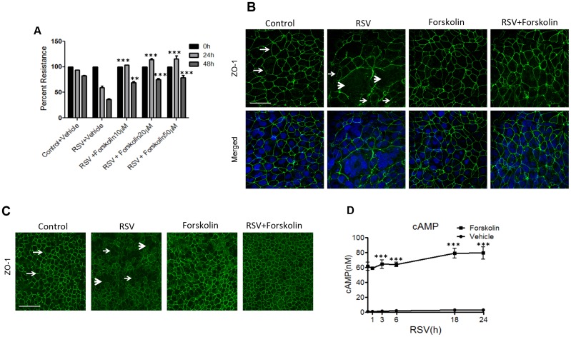
Fig 1. Forskolin attenuates RSV-induced epithelial tight junction disassembly by increasing the intracellular cAMP level.
(A) Confluent epithelial cell monolayers were infected with RSV (MOI, 0.5) for 48 h in the presence or absence of forskolin (10–50 μM,). TEER was measured at indicated time points. (B) Confluent epithelial cell monolayers were infected with RSV (MOI, 0.5) for 48 h in the presence or absence of forskolin (20 μM). Tight junction protein, ZO-1 (green), was visualized by immunofluorescence labeling and confocal microscopy. The nuclei were counterstained with DAPI (blue). Note the characteristic “chicken wire” appearance of ZO-1 in control non-infected cells (arrows), and ZO-1 translocation into cytoplasmic dot-like structures in RSV-infected cells (thin arrowheads). Also, note the syncytia formation in RSV-infected cells (thick arrowheads). Scale bar, 40 μm. (C) Primary human bronchial epithelial cells were infected with RSV (MOI, 2) for 48 h in the presence or absence of forskolin followed by visualizing ZO-1 by immunofluorescence labeling and confocal microscopy. Arrows demonstrate normal junction formation, and thin arrowheads indicate the disappearance of TJ ZO-1 staining in the RSV-infected cell monolayer and thick arrowheads shows syncytia formation. Scale bar, 40 μm (D) Cells were infected with RSV (MOI, 0.5) for 24 h, followed by forskolin treatment (20 μM) for 15 min and subsequent measurement of the cAMP concentration in the total cell lysates. Each image and graph is representative of at least 3 independent experiments. Data is presented as mean ± SEM **, P< 0.01 and ***, P< 0.001 as compared to RSV-infected vehicle-treated cells.

