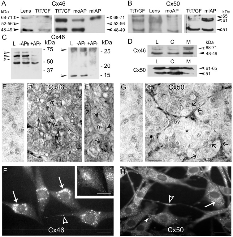
Fig 1. Expression, phosphorylation status and localization of Cx46 and Cx50 in TtT/GF cells and mouse and mink anterior pituitaries.
(A) Left panel: lysates of mouse lens (30 μg) and TtT/GF cells (10 μg), and right panel: lysates of TtT/GF cells (5 μg) and mouse (moAP, 30 μg) and mink (miAP normal male, 20 μg) anterior pituitaries were subjected to SDS-PAGE followed by Western blotting with Cx46 and Cx50 antibodies. Cx46 antibodies detected a 48–49 kDa (arrowhead) band and a 68–71 kDa band (open arrowheads, 71 kDa) in lens. TtT/GF cells, moAP and miAP lysates exhibit the same bands with different intensities. In addition, two faint bands of molecular masses of 52, and 56 kDa were sometimes observed in lens and TtT/GF cells (open arrowheads). (B) Cx50 antibodies revealed a 51 kDa band (arrowhead) and 61 kDa band (open arrowhead) in the lens, TtT/GF cells, moAP and miAP. A third 65 kDa band was detected in TtT/GF cells and in AP lysates (open arrowhead). (C) Cx46 phosphorylation status: TtT/GF cell lysates (control: L) were incubated either in the absence (-APh) or presence (+APh) of alkaline phosphatase. Following incubation, 30 μg protein aliquots from each sample were subjected to SDS-PAGE followed by Western blotting with anti-Cx46. The left panel shows a strong ~68 kDa band flanked by ~71, 56 and 52 kDa bands whose intensities were diminished by the APh treatment (open arrowheads). The right panel shows two Cx46 immunoreactive bands at 25 and 14 kDa in whole cell lysates (L) and in lysates incubated with buffer alone (-APh). The 14 kDa band intensity was reduced by phosphatase treatment (+APh, open arrowhead). (D) TtT/GF cell lysate (L, 30 μg), cytosolic (C, 30 μg) and crude membrane (M, 30 μg) fractions were subjected to SDS-PAGE and immunoblotting with Cx46 and Cx50 antibodies. Representative Western blots show enrichment in the high molecular weight Cx46 and Cx50 immunoreactive bands in the crude membrane fraction (open arrowheads). (E-E”) Cx46 immunohistochemistry in mink AP. (E) No reaction was detected in Cx46 controls done on normal adult male mink APs incubated with either the primary or secondary antibody. (E’) The FS cell delicate cytoplasmic processes were Cx46-positive (arrows). (E”) A higher magnification of a hormone secreting cell labelled with a white asterisk in E’ displays Cx46 labelling (arrowheads) in the perinuclear region and the nucleolus (n). In (E”), the lower arrow points to heavy Cx46 labeling within the cytoplasm of an FS cell; the upper arrows points to Cx46 labelled FS cell thin cytoplasmic process surrounding an adjacent endocrine cell. E and E’, bar: 40 μm. E”, bar: 15 μm. (F) Cx46 immunofluorescence in TtT/GF cells. Cx46 staining showed a punctate, cytoplasmic distribution concentrated in the perinuclear region (arrows). Cx46 labelled cytoplasmic processes (open arrowhead). When changing the focal planes a nuclear distribution became visible (insert). F and insert, bar: 20 μm. (G-G’) Cx50 immunohistochemistry in adult male mink anterior pituitaries. (G) Cx50 controls with either the primary or secondary antibody showed no immunolabeling in adult male mink. (G’) At the periphery of the follicle, the FS cells and their delicate cytoplasmic processes adjoining endocrine cells were Cx50-positive (arrows) in contrast to the hormone secreting cells that were not (asterisk). In addition, Cx50 labeled the wall of blood vessels (bv). G and G’, bar: 40 μm. (H) Cx50 fluorescence microscopy studies in TtT/GF cells. A dust-like Cx50 labelling was evenly distributed in the cytoplasm (arrow). In addition, cytoplasmic processes (open arrowhead) and the cell membrane (arrowhead) were Cx50-positive. No immunoreactivity was apparent in the nucleus. Bar: 20 μm.

