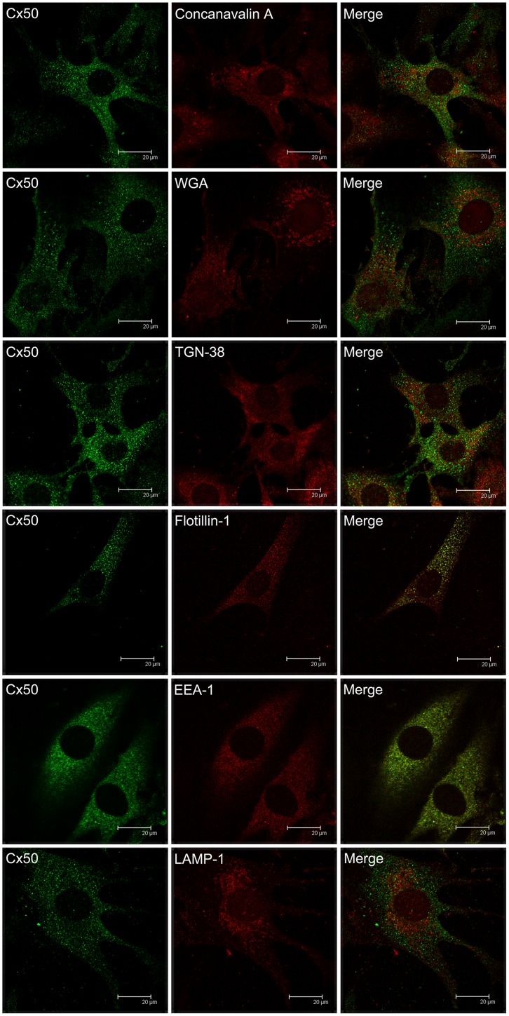
Fig 4. Confocal microscopy on the co-localization of Cx50 and cellular organelles in TtT/GF FS cells.
TtT/GF cells were double-stained with Invitrogen Cx50 antibody and an organelle marker (antibody or probe). Preparations were visualized with a confocal microscope. Micrographs shown correspond to a unique focal plane of 0.7 μm thickness. Cx50 did not co-localize with the endoplasmic reticulum (concanavalin A), Golgi apparatus (WGA), trans-Golgi (TGN-38), early endosomes (EEA-1), or lysosomes (LAMP-1). A weak Cx50 co-localization with the lipid raft marker flotillin-1 was observed in the cytoplasm.

