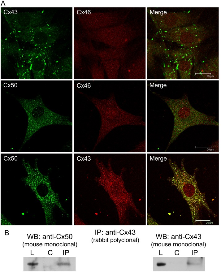
Fig 5. Cx43, Cx46 and Cx50 interaction in TtT/GF FS cells.
(A) Confocal microscopy of TtT/GF cells labelled for Cx43, Cx46 and Cx50. Micrographs shown correspond to a unique focal plane of 0.7 μm thickness. Cx43-Cx46 co-localized neither in the cytoplasm nor in the nucleus. As well, no co-localization was apparent for Cx50-Cx46. Cx50 co-localized with Cx43 in the cytoplasm. (B) Co-immunoprecipitation studies. Pre-cleared TtT/GF cell lysates (L) were incubated either with buffer alone (control: C) or rabbit polyclonal anti-Cx43 (IP). Next, the mixtures were incubated with protein A Sepharose beads. Proteins attached to beads were subjected to SDS-PAGE followed by Western blotting (WB) with either mouse monoclonal anti-Cx50 or mouse monoclonal anti-Cx43. The figure shows representative membranes. Cx43 and Cx50 were both pulled down by Cx43 antibody.

