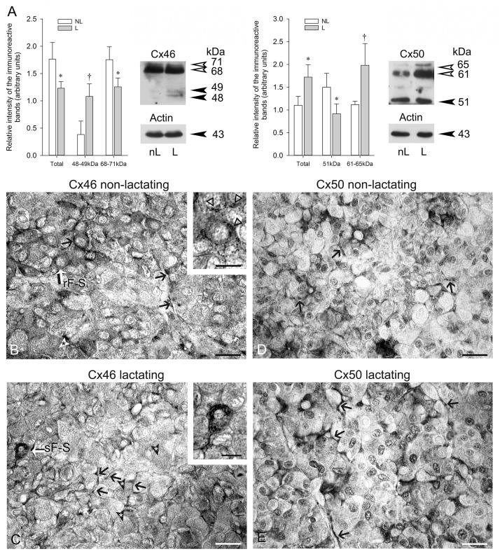
Fig 6. Cx46 and Cx50 in non-lactating and lactating female mink anterior hypophyses.
(A) Western blot analyses of Cx46 and Cx50 in non-lactating (nL) and lactating (L) mink anterior pituitaries. Thirty μg total protein samples were subjected to SDS-PAGE and immunoblotting with Cx46 and Cx50 antibodies. Membranes were reprobed with monoclonal anti-actin and polyclonal anti-actin respectively. Representative Western blots are shown. Values are the mean ± SEM of three different animals per experimental condition. Intensity values were normalized to the corresponding actin value. Statistics (Student’s t test): Total Cx46 levels were lower in lactating than in non-lactating female mink (* P<0.05). The intensity of the 48–49 Cx46 band was higher († P<0.03) whereas that of the 68–71 kDa band was lower (* P<0.05) in lactating than non-lactating mink. Total Cx50 levels were higher in lactating than in non-lactating mink (* P<0.05). The 51kDa Cx50 band intensity was lower (* P<0.05) whereas that of the 61–65 kDa band was higher († P<0.03) in lactating than non-lactating mink. (B-E) Representative light micrographs of non-lactating (B and D) and lactating (C and E) mink anterior pituitary Bouin’s-fixed paraffin sections exposed to Cx46 (B and C) and Cx50 (D and E) antibodies respectively. A Cx46-positive round-shaped type 2 FS cell with a pale rounded nucleus is identified rFS in (B). In addition, open triangles point to Cx46-positive material in the perinuclear Golgi apparatus and lysosomes in three neighboring cells (insert). A type 1 stellate-shaped cell (identified sFS, white arrow) with its nucleus close to the center of the follicle is seen (C) containing plentiful Cx46-positive dots. (B and C): The many arrows indicate Cx46 labelling within the FS cell cytoplasmic processes bordering individual endocrine cells. The white (B) and the black (C) arrowheads with an asterisk point to endocrine cells containing minute Cx46-positive dots. In (D and E), the black arrows point to Cx50-positive FS cell cytoplasmic processes. In (D), a punctate pattern of Cx50 immunolabelling typical of gap junctions is apparent at the site of non-endocrine-endocrine cells contacts associated with plasma membranes. Bars: B, C, D and E: 50 μm; inserts in B and C: 25 μm.

