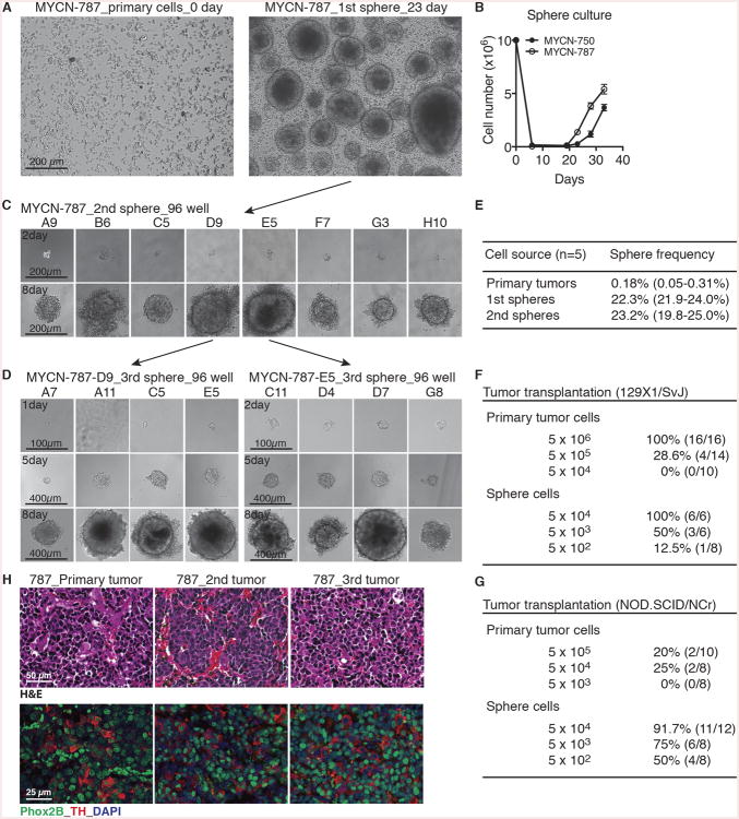
Figure 1. TH-MYCN neuroblastoma sphere-forming cells possess self-renewal and tumorigenic potential.
(A) Phase-contrast images of MYCN-787 primary tumor cells at the day of plating (0 day, left panel) and primary (1st) spheres after culture for 23 days (right panel).
(B) Growth assays of MYCN-750 and -787 primary tumor cells. Spheres appeared after ∼2 weeks of culture. Viable cells were quantified by trypan blue staining. Error bars represent SD (n = 4).
(C-D) Phase-contrast images of serial sphere-forming assays of single cell-derived MYCN-787 secondary (C, 2nd) and tertiary (D, 3rd) spheres.
(E) Quantification of sphere-forming cell frequency in primary tumors, 1st and 2nd spheres.
(F-G) Tumor transplantation assays for primary tumor cells and primary sphere cells in syngeneic 129×1/SvJ (F) a nd NOD.SCID/NCr (G) mice.
(H) Serial tumor transplantation assays with H&E (top panel) and immunofluorescence (bottom panel) staining of MYCN-787 primary tumor, a representative 2nd tumor generated by MYCN-787 primary spheres, and a representative 3rd tumor by spheres from the 2nd tumor. Nuclei were stained with DAPI.
See also Figures S1.

