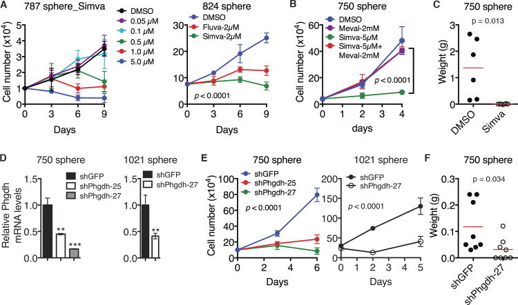
Figure 4. Cholesterol and serine-glycine pathways are essential for maintaining neuroblastoma sphere-forming cells.
(A) Growth assays of neuroblastoma sphere-forming cells treated with DMSO, increasing concentrations of Simvastatin (Simva) or 2 μM Fluvastatin (Fluva). Error bars represent SD (n = 4). Data were analyzed by two-way ANOVA with p values indicated.
(B) Growth assays of neuroblastoma sphere-forming cells treated with DMSO and Simva with or without mevalonate (Meval). Error bars represent SD (n = 4). Data were analyzed by two-way ANOVA with the p value indicated.
(C) Tumor transplantation assay of neuroblastoma sphere-forming cells treated with DMSO or 5 μM Simva for 24 hours. Tumor weight was analyzed by scatter plot with horizontal lines indicating the mean.
(D) qRT-PCR analysis of Phgdh mRNA levels in neuroblastoma sphere-forming cells infected with lentiviruses expressing shRNA against GFP or Phgdh. Error bars represent SD (n = 3).
(E) Growth assays of neuroblastoma sphere-forming cells without or with Phgdh knockdown. Error bars represent SD (n = 4). Data were analyzed by two-way ANOVA with p values indicated.
(F) Tumor transplantation assay of neuroblastoma sphere-forming cells without or with Phgdh knockdown. Tumor weight was analyzed by scatter plot with horizontal lines indicating the mean.
**p < 0.01, ***p < 0.001. See also Figure S4.

