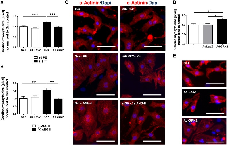
Fig 3. G-protein coupled receptor kinase 2 (GRK2) promotes cardiac myocyte hypertrophy.
(A-C) GRK2 knockdown by siGRK2 attenuates G-protein coupled receptor (GPCR) induced cardiac hypertrophy. (A) Quantification of cardiac myocyte cell size from immunofluorescence stains of neonatal rat ventricular myocytes (NRVMs) following siRNA mediated GRK2 knockdown and GPCR stimulation by phenylephrine (PE), >200 NRVM per condition from 3 independent cell preparations were analyzed, *** P < 0.001. (B) Quantification of cardiac myocyte cell size following siRNA mediated GRK2 knockdown and Angiotensin II (ANG II) treatment, >100 NRVM per condition from 3 independent cell preparations were analyzed, **, P < 0.01. (C) Representative immunofluorescence images of NRVMs under respective conditions. α-Actinin is stained red for demarcation of cell dimensions, blue DAPI staining marks nuclei. (D, E) GRK2 overexpression promotes hypertrophy. (D) Quantification of cardiac myocyte cell size from immunfluorescence stains of NRVMs following treatment with an adenovirus harboring GRK2 (AdGRK2) or β-galactosidase/LacZ as control (AdLacZ), >100 NRVM per condition from 3 independent cell preparations were analyzed, *, P < 0.05. (E) Representative images of immunofluorescence of NRVMs (DAPI and α-Actinin) following AdGRK2 or AdLacZ transduction compared or untreated (-) cells. Scale bar (C and E): 50 μm.

