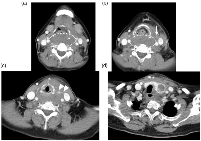
Fig. 1.
Initial enhanced CT. A large hematoma was observed in the retropharyngeal space (dashed oval (a, b)). The left superior thyroid artery is clearly depicted (arrow in (b)), which connects to an extravasation (arrowheads in (c)). The left lobe of the thyroid gland was enlarged and contained unenhanced low-density areas (d). Hematomas were also observed around the left lobe of the thyroid and in the upper mediastinum (d).

