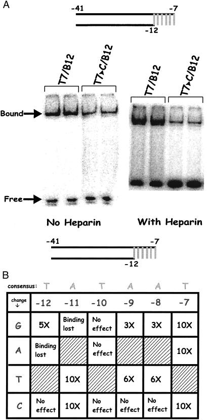
Figure 5.
EMSA on fork + tail probes in the presence of heparin. (A) EMSA comparison of probes with changes at −7 in the presence and absence of heparin (Right panel is overexposed to show low binding). (B) Data summary for all substitutions in the presence of heparin, as described in Fig. 4.

