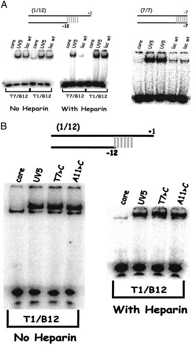
Figure 6.
EMSA on lac wild type and UV5 by using probes that reach the start site. The UV5 mutation is a double change (−8T and −9G). (A Left) Comparative data for the longer 1/12 and shorter 7/12 probes. Lac wild-type binding is within a factor of two of the UV5 binding except on T7/B12 in the presence of heparin, which differs by a factor of 10. (Right) This panel is overexposed to show low binding; wild-type binding differs by a factor of 10 on duplex probe (7/7), but this difference is eliminated by addition of base pairs to + 1 (data not shown). (B) On the longer 1/12 probes, substitutions in important positions within the consensus have little effect.

