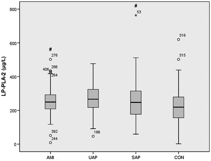
Figure 1.
Box-whisker plots showing levels of Lp-PLA2 (µg/l) in Chinese patients who underwent coronary angiography and were diagnosed with coronary heart disease, divided into patients with acute myocardial infarction (AMI; n = 72), unstable angina pectoris (UAP; n = 254) or stable angina pectoris (SAP; n = 65), compared with a control group of patients with normal coronary angiography (CON; n = 140). #P < 0.05 versus controls. Central black horizontal line within the box, median; box extremities, upper and lower-quartiles; error bars, 1.5 times the interquartile range; ○, mild outlier; and *, extreme outlier

