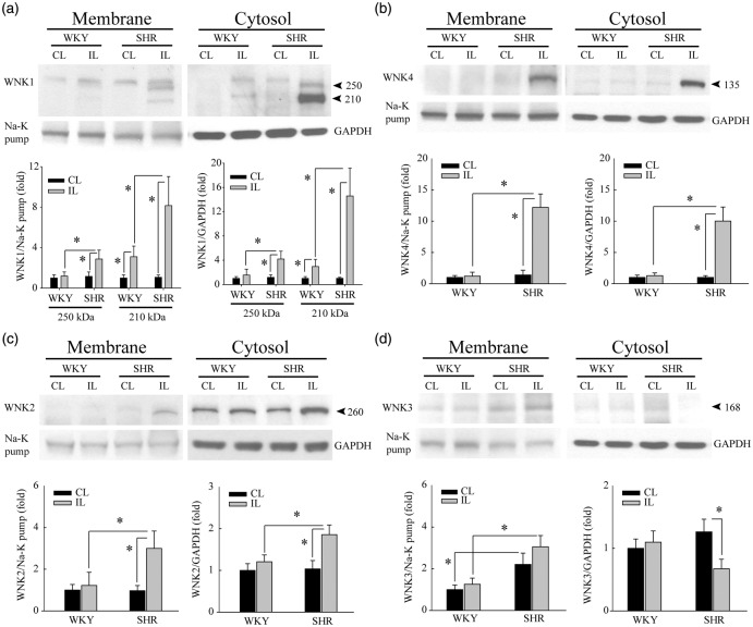
Figure 3.
SHR brains exhibit pronounced upregulation of WNK1, WNK2, and WNK4 proteins after ischemic stroke. (a) Upregulation of full length WNK1 protein (250 kDa) was detected at 6-h reperfusion post-tMCAO only in IL cortices of SHR but not WKY rats. Cytosol and crude membrane protein fractions were prepared from contralateral (CL) and ipsilateral (IL) cortices of WKY and SHR. Data are mean ± SD, n = 4, *p < 0.05. (b) Upregulation of WNK4 protein in the same samples as in (a). Data are mean ± SD, n = 4, *p < 0.05. (c) Upregulation of WNK2 protein in the same samples as in (a). Data are mean ± SD, n = 4, *p < 0.05. (d) Ischemic stroke did not cause significant changes of WNK3 protein expression in either WKY or SHR brains but reduced WNK3 expression in the IL cytosol fraction of SHR brains. Data are mean ± SD, n = 4, *p < 0.05.

