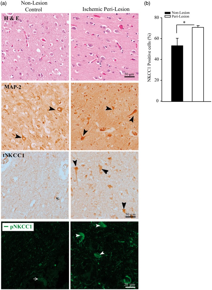
Figure 6.
Elevated tNKCC1 and pNKCC1 protein expression in subacute human middle cerebral artery ischemic stroke brain autopsy. (a) Non-lesion and peri-lesion regions in human ischemic stroke brain autopsy samples were identified with H & E staining. tNKCC1 expression in neuronal cells was shown in representative immunohistochemical images. Arrow: low NKCC1 expression. Arrowhead: high NKCC1 expression. Scale bar: 50 µm. Representative immunofluorescence images of pNKCC1 expression in brain cells (bottom panel). Arrow: low pNKCC1 expression. Arrowhead: high pNKCC1 expression. Scale bar: 20 µm. (b) Percentage of tNKCC1-positive cells among total To-pro3-positive cells. Numerical data are mean ± SD, n = 3 (one male, two female), *p < 0.05.

