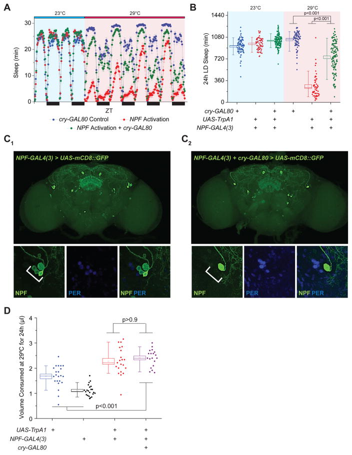
Figure 3. A Subset of Circadian Pacemaker Cells Largely Regulates NPF Effects on Wakefulness but Not Feeding.
(A–B) Repressing activation of NPF signaling in cry-expressing cells largely revert the sleep loss phenotype, illustrated as (A) a sleep profile and (B) as a box plots quantifying total sleep amount. n=59–128. (C) NPF-GAL4, cry-GAL80 expression pattern reveals NPF expression in a subset of the PER+ LNd circadian pacemaker neurons. (Top) Representative expression pattern of adult brain images for (C1) NPF-GAL4 and (C2) NPF-GAL4, cry-GAL80 using membrane GFP as a reporter. White arrows denote location of LNd neurons in the adult brain. (Bottom) High magnification images of region indicated by white arrows in top panels demonstrating co-localization of NPF (green) and PER (blue) within LNd neurons. White scale bars in images denote 25 μm. (D) Repressing activation of NPF signaling in cry-expressing cells does not alter adult feeding behavior. n=19–21 experimental replicates. All error bars indicate SEM. See also Figure S4.

